Scanning Electron Microscopy (SEM) of PMMA Nanospheres
and Inverse Opal Photonic Crystals
Samples are deposited on conducting graphite tape and gold coated.
The coated samples are placed into an electron microscope.
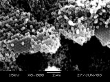
Zooming in on a sample of monodispersed polymethylmethacrylate spheres.
What is the diameter of the spheres?
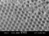
Zooming in on a sample of inverse opal photonic silica crystal. What
is the diameter of the holes?
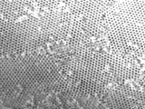
Flying over the surface of a sample of inverse opal photonic crystal
(2000x magnification.)
Developed in collaboration with the
University of Wisconsin Materials Research Science and Engineering Center
Interdisciplinary Education Group | MRSEC on Nanostructured Interfaces
This page created by George Lisensky, Beloit College. Last modified June 16, 2013 .
University of Wisconsin Materials Research Science and Engineering Center
Interdisciplinary Education Group | MRSEC on Nanostructured Interfaces
This page created by George Lisensky, Beloit College. Last modified June 16, 2013 .


