Suspension polymerization and size determination of monodispersed polymethylmethacrylate (PMMA) nanospheres
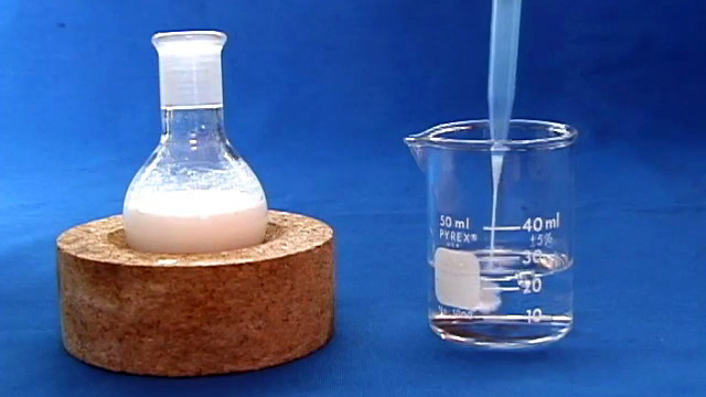
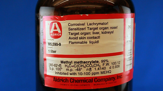
Monodispersed polymethylmethacrylate nanospheres are synthesized from the suspension polymerization of a rapidly stirred aqueous suspension of methyl methacrylate. 2,2-Azobis(2-methylpropionamidine) dihydrochloride is a heat activated water soluble free radical initiator so the emulsion droplets polymerize starting from their outer edge.
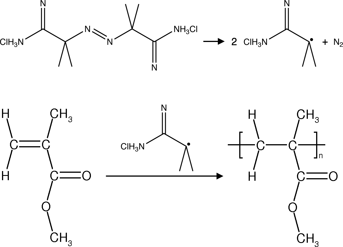
The small uniform diameter particles should appear irridescent if they are close-packed and their size is similar to the wavelength of visible light. The size of the polymethylmethacrylate spheres can be characterized using the settling velocity and Stoke's Law, using evaporative self-assembly and visible spectroscopy, and direct measurement using scanning electron microscopy. The size can also be measured after using the spheres as a template and creating a silica inverse opal.
| Procedure | Wear eye protection |
Chemical gloves recommended |
 Fumehood required Fumehood required |
Synthesis (Write a purpose and method for the synthesis before starting. What are the important variables?)
Procedure modified by George Lisensky, Jacob Horger, and Jiaqi Luo, Beloit
College, from the
Inverse Opal Photonic Crystals Laboratory Guide by R. Schroden and
N. Balakrishnan, University of Minnesota MRSEC, 2001.
(Not using a teflon sleeve usually results in a permanently polymethylmethacrylate fused joint at the end of the experiment.)
(It is important to use a stir bar designed for a round bottom flask for maximum stirring.)
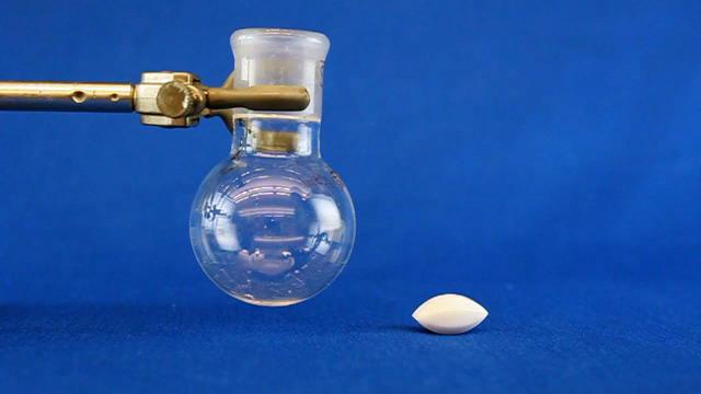
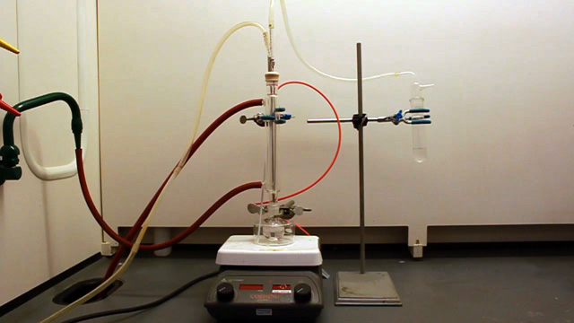
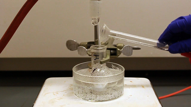
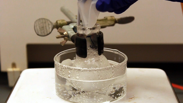
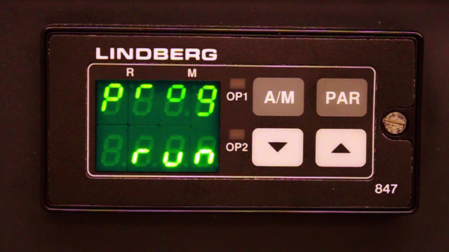
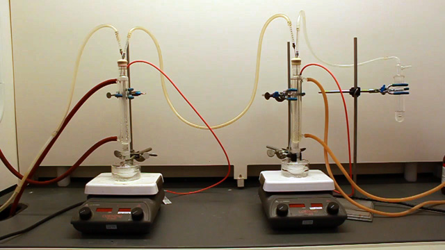
Connect the water lines to the condenser (in at the bottom and out at the top). Put water in the crystallizing dish and heat to 70 °C. (An automatic temperature controller may be available. The crystallizing dish containing water helps minimize temperature fluctuations and provides a place to measure the temperature.) Spin the stir bar at the maximum speed that works. What is your stir speed?
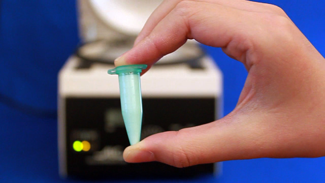
 Click image for larger view
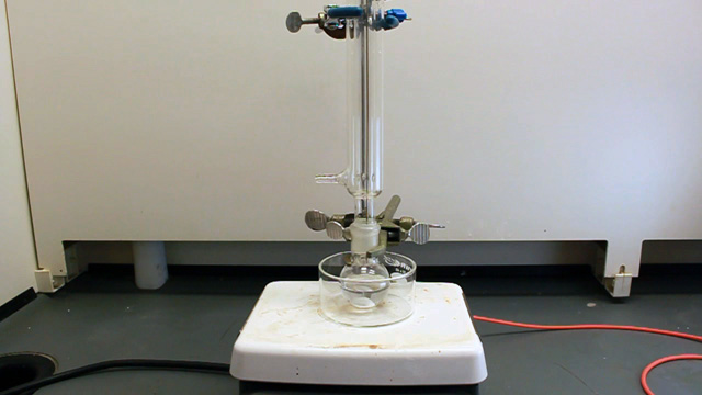
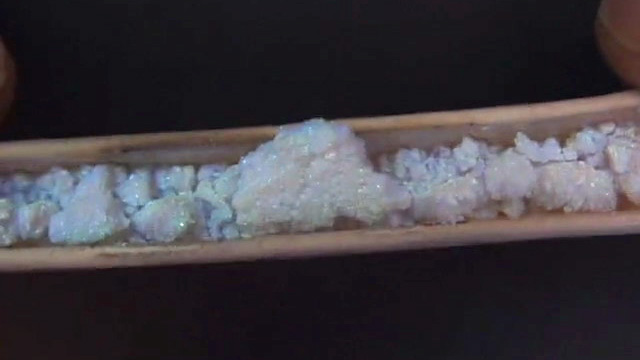
Product without nitrogen
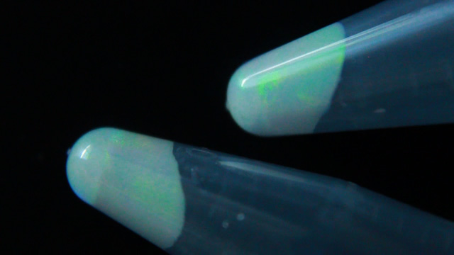
 Click image for larger view
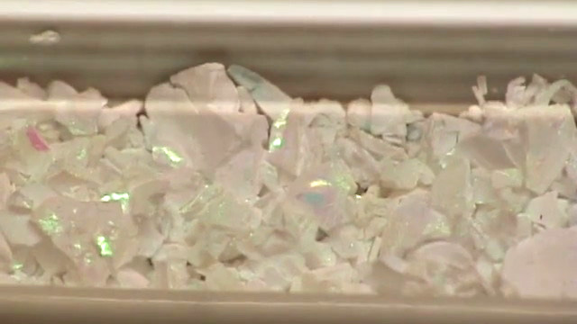
Poorly stirred product
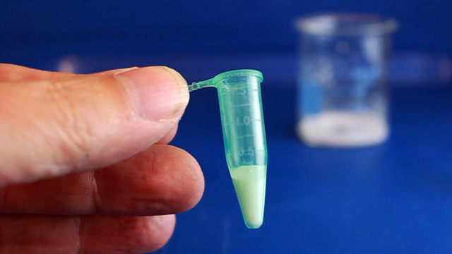
 Click image for larger view
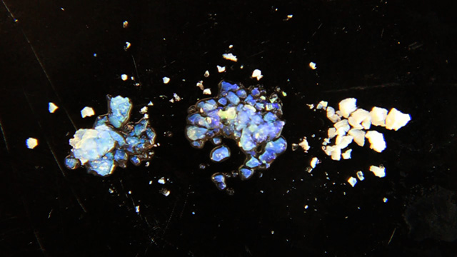
Well stirred product
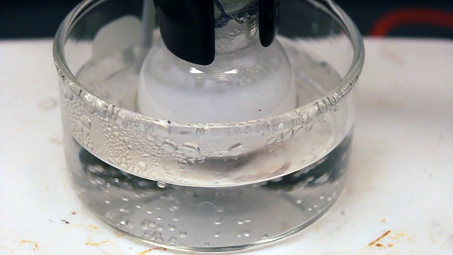
After 40 minutes of heating, remove the condenser. There should not be a noticeable odor if the polymerization was successful. Be sure to return the Teflon joint sleeve.
After the synthesis (same lab period)
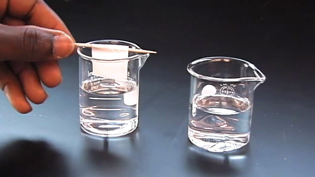
- Use the original solution to set up the Stokes Law measurements.
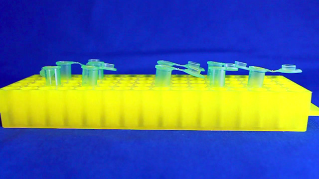
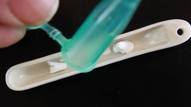
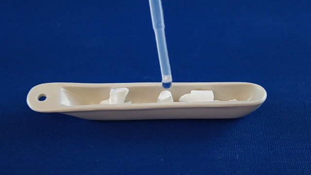
- Add 500µL of original solution to 20 mL water in a large centrifuge tube. Save the capped tube to begin diffraction and SEM experiments next week.
- Use the original solution to fill four labeled microcentrifuge tubes. Save the capped tubes to begin inverse opal synthesis experiments next week. Ideally these tubes will be centrifuged at 2000 rpm for several hours before the next lab period.
- Store any remaining original solution in a capped appropriate-size centrifuge tube for insurance.
- Glassware can be cleaned with acetone since acetone dissolves the starting material and the product.
Stoke's Law Determination of Particle Size (Write a purpose and method for the Stoke's Law determination before starting.)
Procedure developed by George Lisensky and Jiaqi Luo, Beloit College.
Either print each photograph bigger than life on a full sheet of paper and use a ruler or use ImageJ for measuring lengths in digital images. (Use the straight line tool to drag the length of the black tape and menu Analyze/Set Scale to tell the program the actual length of the tape, often 19mm. You can then use the straight line tool to measure lengths in real units.) Be sure all measured data is recorded in your laboratory notebook.
Calculations and Graphs
The beads will fall at the terminal velocity, which depends on their size and the known viscoscity and densities.
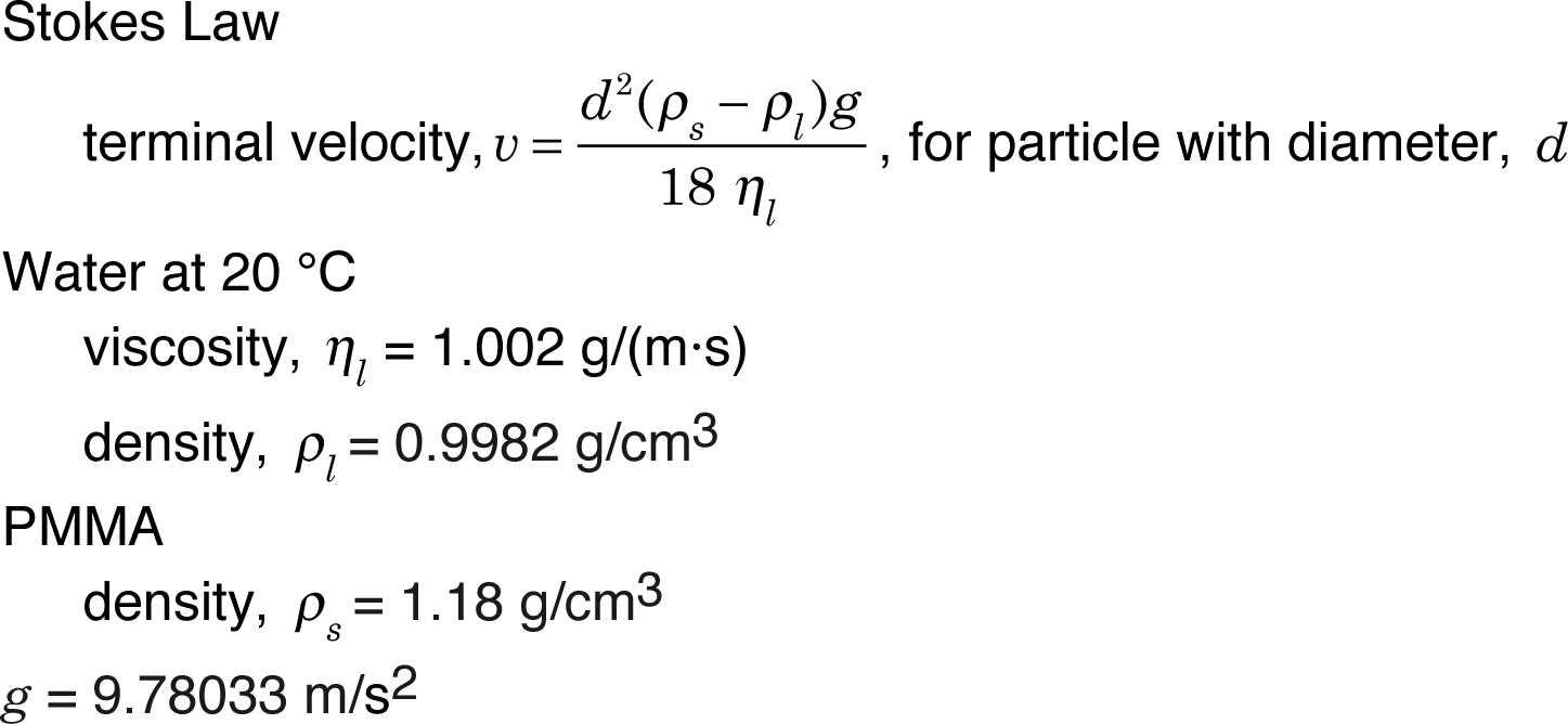
Update your data table at least weekly. For each data point (total time in seconds, distance fallen in mm), calculate the particle diameter. Is the calculated diameter consistent?
Near the end of the experiment plot the distance fallen in mm as a function of the number of seconds and use the slope of the linear portion as the best fit velocity. What is the overall velocity in nm/s? What is the particle size based on this velocity? What does it mean if your y-intercept is not zero?
Optical Diffraction Determination of Particle Size (Write a purpose and method for the diffraction experiment before starting. What are the important variables?)
Procedure by William Schreiter, Richard Amankwah and Karen Nordell, Chemistry Department, Lawrence University.
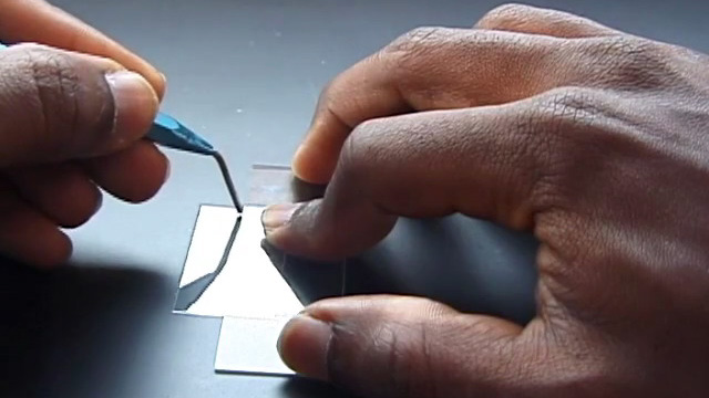
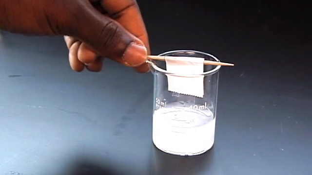
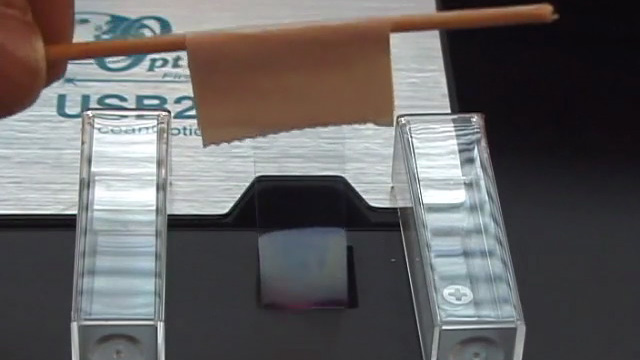
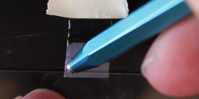
Evaporation of a dilute suspension of polymer nanospheres creates a close packed layer on the surface of a glass slide suspended in the solution. The particle size can be calculated from the visible absorption maximum of this colloidal crystal.
 Click image for larger view
25 watt incandescent lamp
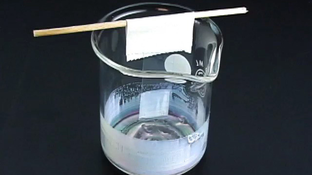
Observe the color of the film under reflected light conditions by holding the slide under a bright light source. Observe the color of light transmitted through the film by holding the glass slide up and looking at a bright light source through the slide. How are the colors of reflected and transmitted light related?
Use a modified Bragg equation to calculate the size of the nanoparticles from the wavelength of the absorbance peak, λp. The index of refraction is 1.49 for PMMA and 1.00 for air.
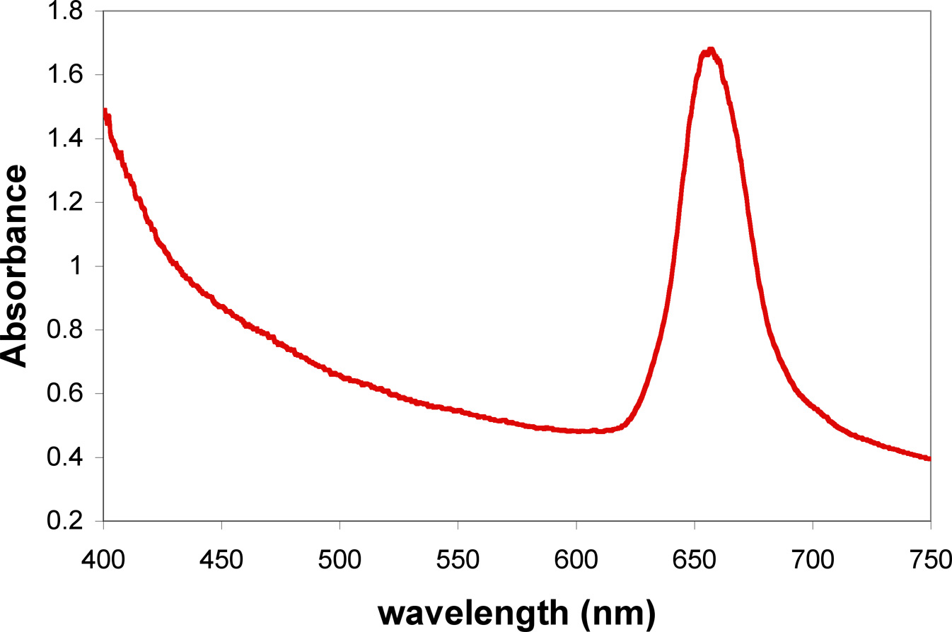
λp = the wavelength at maximum absorbance (in nm)
Ds = the diameter of the close packed spheres (in nm)
ns = the index of refraction for the spheres
nv = the index of refraction for the void spaces (presumably air in a dry film)
f = the filling fraction for a close packed structure (0.76)
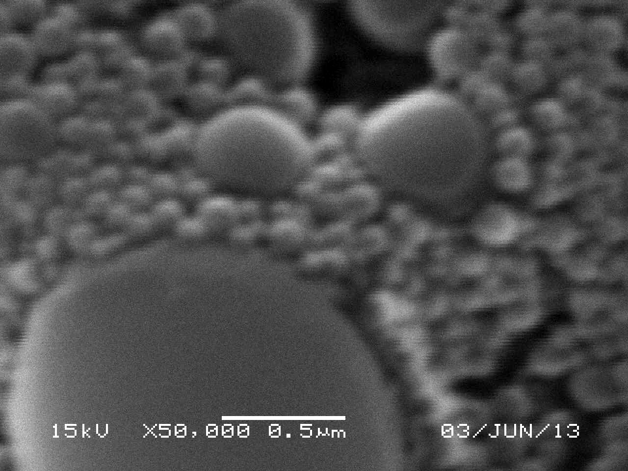
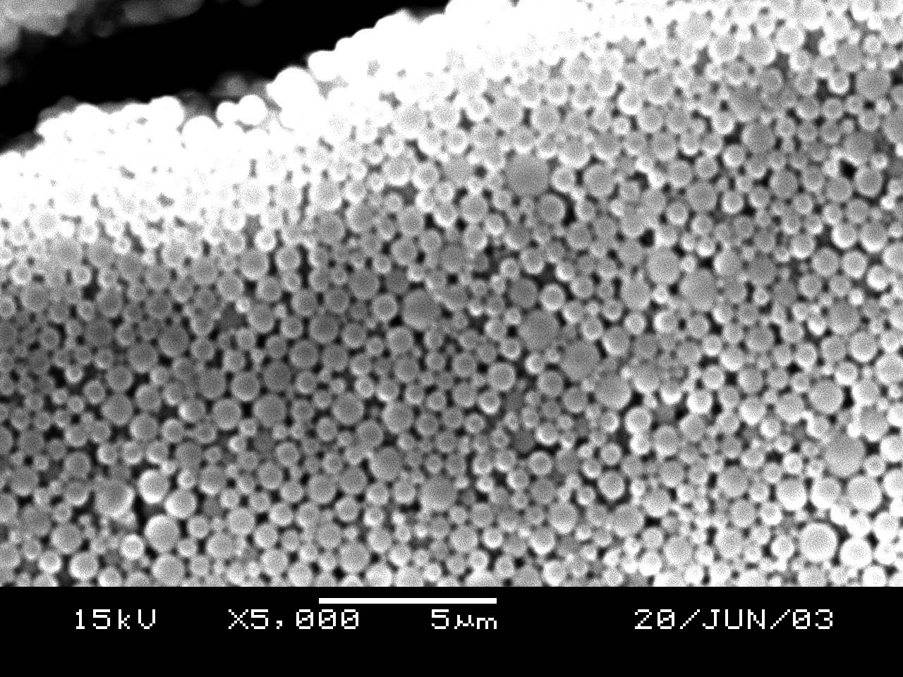
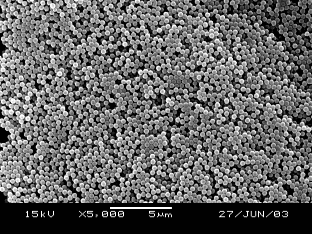
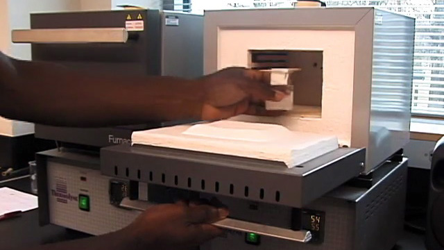
SEM Determination of Particle Size (uses sample from previous step)
Synthesis of Inverse Opal Photonic Crystals (Write a purpose and method for the synthesis before starting.)
Procedure modified by George Lisensky, Jacob Horger and Jiaqi Luo, Beloit
College, from the
Inverse Opal Photonic Crystals Laboratory Guide by R. Schroden and
N. Balakrishnan, University of Minnesota MRSEC, 2001.
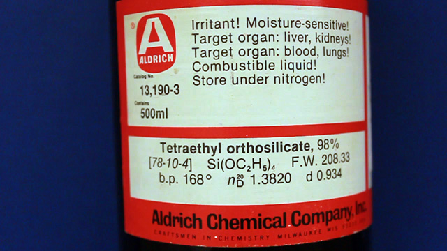
A sol-gel synthesis using tetraethylorthosilicate and close-packed polymethylmethacrylate spheres as a template yields a dimensionally ordered porous silica solid.
SEM Determination of Inverse Opal Hole Size (uses sample from previous step)
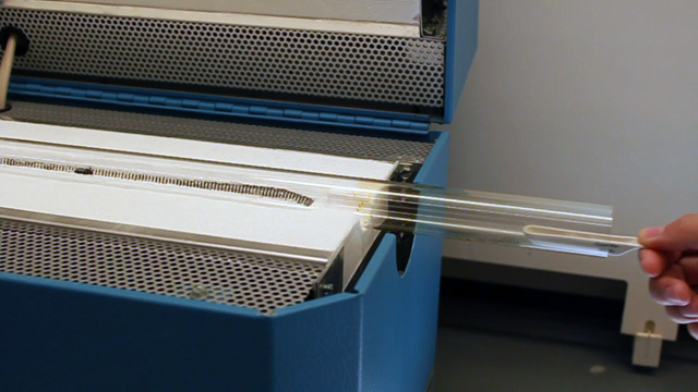
Stick a portion of your sample to the carbon tape on the SEM holder and add your name to the sample map. Samples will then need to be sputtered since SiO2 is non-conducting. On our instrument (using 25kV with spotsize 27 and z 15mm) it should be possible to see holes at 3000x. Look carefully. Hole size is better measured using multiple holes in a line and then dividing by the number of holes measured. (ImageJ is useful for measuring lengths. Use the straight line tool to drag the length of the scale bar and menu Analyze/Set Scale to tell the program the actual length of the scale bar. You can then use the straight line tool to measure lengths in real units.) Be sure to include details of your analysis in your laboratory notebook.
Inverse Opal Properties (uses sample from previous step)
- What color is your product in air? (The index of refraction of silica is 1.460 and of air is 1.000)
- Add a drop of ethanol to a small portion of your product. What color do you observe? (The index of refraction of ethanol is 1.360)
- Add a drop of toluene to a small portion of your product. What color do you observe? (The index of refraction of toluene is 1.496)
- Based on your SEM data, predict the absorption wavelength for air, ethanol, and toluene. How do you need to modify the equation for closed-packed spheres for the inverse material? How well do your predictions match the observed colors? Explain.
Spectra, Images and Graphs
Your notebook should include your Stoke's Law photographs and graph, optical diffraction spectra, and SEM images of both opal and inverse opal materials.
- Did your methyl methacrylate polymerize? What observations support your answer? Using your chemical knowledge of polar/nonpolar, physical properties, and the polymerization mechanism, explain why solid spheres are expected to be produced.
- Based on your Stoke's Law measurements, what is the velocity (nanometers/second) and the corresponding diameter (nanometers) of the spheres?
- Based on evaporative self-assembly to pack spheres, what is value for the photonic band gap (nanometers or eV) and the corresponding diameter (nanometers) of the spheres? What did you use to obtain a constant temperature? How many times did you try the evaporation?
- Based on the SEM image of the opal, did the spheres close-pack and what is the diameter of the spheres (nanometers)? You should measure the diameter for more than one sphere and more than one image and take an average. Report all measured data.
- Based on the SEM image of the inverse opal, are the holes close-packed and what is the diameter of the holes (nanometers)? You should measure the diameter for more than one hole and more than one image and take an average. Report all measured data.
- Based on your hole diameter, predict the absorption wavelength for air, ethanol, and toluene. (How do you need to modify the equation for closed-packed spheres for the inverse material?) How well do your predictions match the observed colors?
- What size are the nanospheres? Do the different methods give the same results within experimental error?
Materials for Synthesis and Stokes Law
Materials for Diffraction
Materials for Inverse Opal
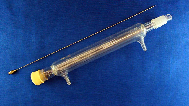
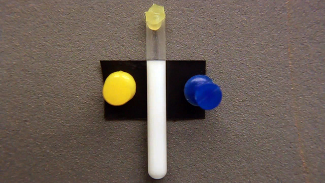
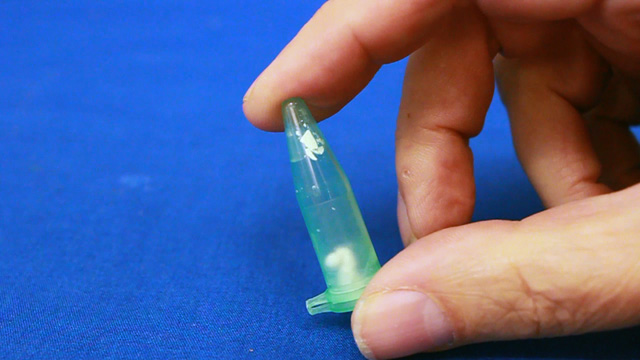
 Click image for larger view
Use a tube as shown on right.
University of Wisconsin Materials Research Science and Engineering Center
Interdisciplinary Education Group | MRSEC on Nanostructured Interfaces
This page created by George Lisensky, Beloit College. Last modified May 15, 2019 .