Procedure from
A. K. Bentley, M. Farhoud, A. B. Ellis, G. C. Lisensky, Anne-Marie Nickel, and W. C. Crone,
"Template Synthesis and Magnetic Manipulation of Nickel Nanowires,"
Journal of Chemical Education, 82,
765-768 (2005). Thanks to Anupam Ghosh for suggesting option C. This version of the experiment uses electrical tape to hold the filter.
It uses less equipment but taping the filters without breaking them requires careful manipulation.
Another version uses a syringe holder, clamps, and an o-ring instead of taping.
A simple way to make nanowires is
to use a mold or template. In this experiment nickel nanowires are grown inside
the pores of an alumina filter and then the filter is removed by etching to
yield magnetic nanowires.
Nanoporous membranes were designed for health care applications
including virus filtration, sample preparation, and liposome manufacture (http://www.whatman.com).
These alumina membranes are manufactured by applying a large electrical potential
to a piece of aluminum metal submerged in an acid. Aluminum is oxidized to
alumina (Al2O3) and pores are created. The size of the
pores depends on the applied potential.
Avoid contact with or inhalation of nickel and nickel solutions.
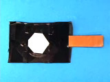
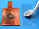
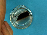
Obtain a 0.02 micrometer Anodisc filter. These ceramic discs are
quite brittle and are supported by a polymer ring. Always use tweezers
to hold the membranes by the support ring; the alumina will crack if handled
directly. Remove the disc from the packaging, remembering which side was
up in the box. Fully coat the upper side (the polymer ring looks wider)
with a conducting metal (see options in next steps).
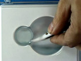
One option is to use a cotton applicator and liquid GaIn alloy to
paint the surface. The coated side will look shiny (and the opposite side
will remain lighter.) While it is important to fully coat the surface
to prevent leaks, it is only necessary to dip the applicator in the GaIn
once. The GaIn can be spread quite thin. Check for gaps in the GaIn coating
by looking at the non-coated face of the membrane. Any areas without GaIn
will appear light blue in color while areas with GaIn will appear white
or opaque.
Another option is to sputter Ag metal onto the surface. Conditions
used were 50 millitorr argon, 45 milliamps current, for three 150 second
depositions. The coated side will look shiny (and the opposite side will
remain lighter.)
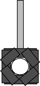
Use electrical tape to fasten the disc, metal coated side down,
to a copper electrode. Attach all sides of the disc so solution
cannot leak behind the disc. Cover the bottom and back portion of
the copper electrode so it won’t come in contact with the
electrolysis solution.
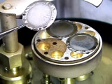
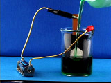
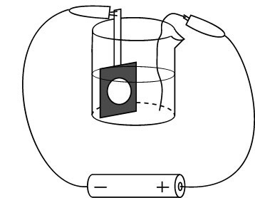
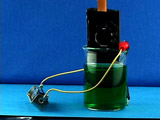
Connect the negative lead of a 1.5 V battery to the copper electrode
and the positive lead to a nickel wire in a 50 mL beaker. Add nickel
plating solution to cover the disc. Electrolyze for 10-50 minutes.
Longer times give longer wires.
Disconnect the battery and remove the copper electrode from solution.
The nickel solution can be reused for this experiment.Why does the concentration
of nickel in solution not change during the electrolysis?
Rinse the electrode with water.
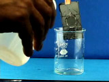
Immerse the electrode in acetone to remove the adhesive. The tape
will easily come off the electrode in a few miniutes. (The second movie
is time lapse photography and represents 15 minutes actual time.)
Remove the disc from the copper electrode and tape shiny side up to
a glass slide for removal of the metal coating.
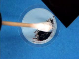
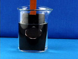
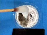
In a fume hood, use concentrated nitric acid and a cotton applicator
to remove the shiny GaIn or the Ag coating. Soak the cotton applicator
in water before disposal.
Rinse with water.
Option A: Obtain the powder x-ray diffraction spectrum (2θ = 40-100°) of the nickel nanowires in the filter.
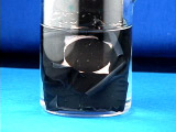
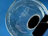
Place the disc in 5 mL of 6 M NaOH for 10 minutes. The ceramic material
will dissolve. Discard the polymer support ring.
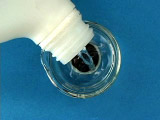
Place the beaker on a strong magnet. The nickel nanorods will be attracted
towards the magnet. Remove the NaOH solution. Add water to rinse, place
the beaker on a strong magnet, and remove the rinse solution. Repeat several
times. Transfer the final suspension to a vial for storage. Keep the wires
in solution.
Option A: Obtain the powder x-ray diffraction spectrum (2θ = 40-100°) of the free nickel nanowires.
Option B: Use an SEM to measure the length of the nanowires. Does the length correlate with deposition time?
Option C: Rinsing with ethanol (and storing the nanowires in ethanol) gives a suspension that leaves less residue and that evaporates more quickly for preparation of SEM samples.
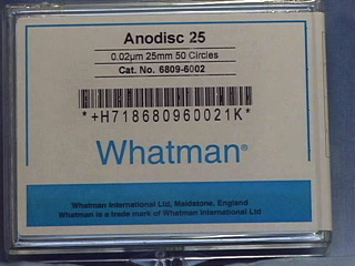
Materials
Whatman Anodisc alumina filters 25 mm with polypropylene support rings and
0.02 micrometer pores (not available directly from Whatman):
Fisher Scientific (#09-926-34) or VWR International (#28138-067)
GaIn Eutectic: Aldrich (49542-5)
"Watts" Nickel plating solution: 300g/L NiSO4.6H2O, 45g/L each H3BO3
and NiCl2.6H2O.
Ni wire: VWR International (#AA41361-G6) or Alfa Aesar 1 mm diameter x 10 m long. CAUTION: Avoid physical contact (especially inhalation) with nickel and
nickel solutions as nickel is an irritant and carcinogen.
Copper sheet: 0.032 inch thick satin finish copper sheet, 12" x 12", McMaster-Carr (#9801K11) http://www.mcmaster.com
Tweezers, cotton swabs, electrical tape, scissors, water wash bottle, 50 mL beaker
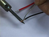
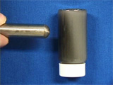
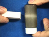
The movie shows assembly of the battery holders. Cut in half a wire with
aligator clips on each end. Strip off the insulation for a short distance.
Use a soldering iron to melt some solder onto each wire and onto the ends
of the battery holder, then use a soldering iron to connect the wires
to the battery holder.
Properties
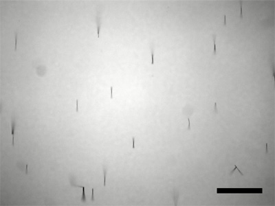
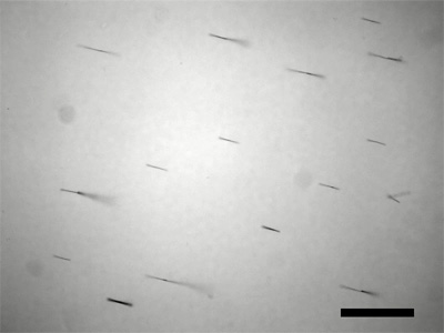
Nickel nanowires viewed through an optical microscope (20x) while
a magnet is moved back and forth from the front to the side of the microscope.
The nanowires rotate to align with the magnetic field. The scale bar represents
100 micrometers.
Nickel nanowires viewed through an optical microscope (20x) while
a magnet is spun at one side. The nanowires rotate to align with the magnetic
field. The scale bar represents 100 micrometers.
Nickel nanowires suspended in water controlled by a magnetic field.