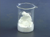
Synthesis of Nanocrystalline Y2O3:Eu Phosphor
Procedure based on D. B. Bolstad and A. L. Diaz, J. Chem. Educ., 79, 1101-1104 (2002).
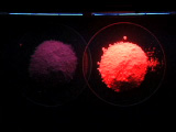
A small percentage of cations in an inert Y2O3 host are replaced by luminescent Eu+3 ions to give a red phosphor. The quality of x-ray diffraction depends on the particle size and the efficiency of the 4f phosphor transitions is also influenced by crystallinity of the host. In this experiment measurements are made on the initial product and on the product after further heating. The study of solid-state luminescence impacts a wide variety of technologies, including display (CRTs and flat televisions), lighting (fluorescent lamps and mercury-free lamps), and medical imaging.
| Procedure | Wear eye protection |
Chemical gloves recommended |
 Fumehood recommended |
Thermal gloves recommended |
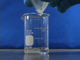
Cover with a watch glass.
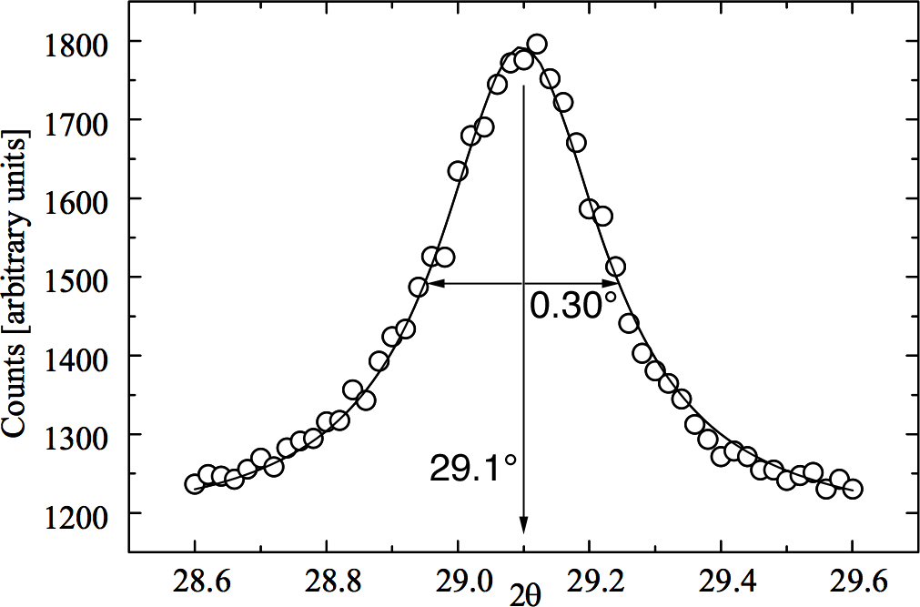
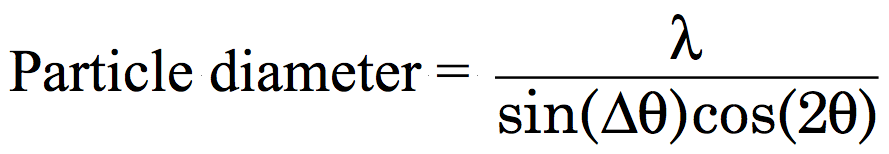 where λ is the X-ray wavelength, Δθ is the peak-width at half-height (FWHM) and 2θ is the peak location. (For even better results use the peak width of the sample minus the peak width of the same peak in a bulk sample as the peak width.)
The X-ray wavelength depends on the target in the instrument X-ray tube. Common targets are Cr (2.28970Å), Fe (1.93604Å), Co (1.78897Å), Cu (1.54056Å), or Mo (0.70930Å) metals.
where λ is the X-ray wavelength, Δθ is the peak-width at half-height (FWHM) and 2θ is the peak location. (For even better results use the peak width of the sample minus the peak width of the same peak in a bulk sample as the peak width.)
The X-ray wavelength depends on the target in the instrument X-ray tube. Common targets are Cr (2.28970Å), Fe (1.93604Å), Co (1.78897Å), Cu (1.54056Å), or Mo (0.70930Å) metals.
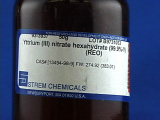
- Yttrium(III) nitrate hexahydrate(99.9%), Strem 93-3937 or Aldrich 237957-25G
- Europium(III) nitrate hexahydrate (99.9%), Strem 93-6310 or pentahydrate Aldrich 207918-1G
- Urea
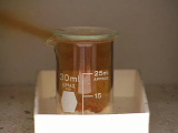
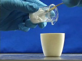
- 30 mL beaker and watch glass
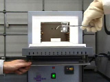
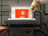
- 500 degree C oven and tongs
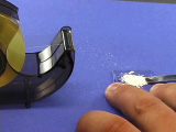
- crucible and cover
- 850 degree C oven
- ultraviolet light
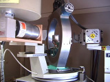
- powder diffractometer
Equipment
Developed in collaboration with the
University of Wisconsin Materials Research Science and Engineering Center
Interdisciplinary Education Group | MRSEC on Nanostructured Interfaces
This page created by George Lisensky, Beloit College. Last modified May 13, 2019 .
University of Wisconsin Materials Research Science and Engineering Center
Interdisciplinary Education Group | MRSEC on Nanostructured Interfaces
This page created by George Lisensky, Beloit College. Last modified May 13, 2019 .