Emulsion polymerization, size determination, and self-assembly of monodispersed polymethylmethacrylate (PMMA) nanospheres for photonics
George Lisensky, Fabian Dauzvardis, Jiaqi Luo, Jacob Horger, and Emma Koenig, Beloit College
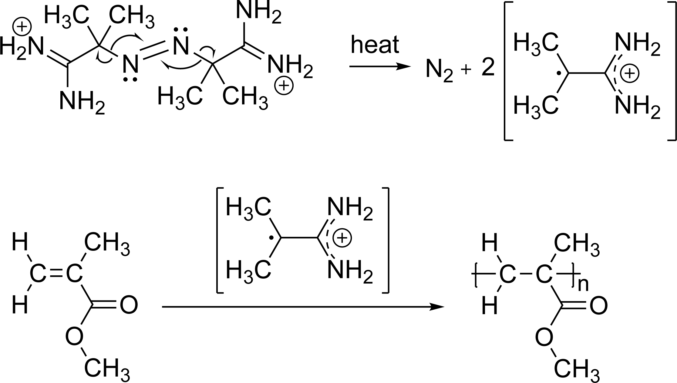
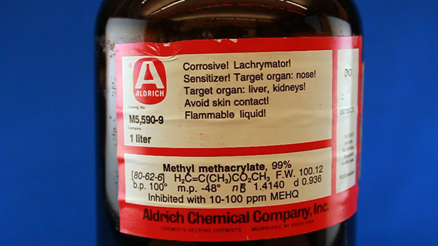
Monodispersed polymethylmethacrylate nanospheres are synthesized from the emulsion polymerization of a rapidly stirred aqueous suspension of methyl methacrylate.
2,2-Azobis(2-methylpropionamidine) dihydrochloride is a heat activated water soluble free radical initiator so the emulsion droplets polymerize starting from their outer edge.
The small, uniform diameter particles should appear irridescent if they are close-packed and their size is similar to the wavelength of visible light.
The size of the polymethylmethacrylate spheres can be characterized using the settling velocity and Stoke's Law, using evaporative self-assembly and visible spectroscopy measurement of the photonic band gap,
and direct measurement using scanning electron microscopy. The size can also be measured after using the spheres as a template and creating a silica inverse opal.
Synthesis (Write a purpose and method for the synthesis before starting. What are the important variables? What is the chemical reaction?) This procedure is derived from the Inverse Opal Photonic Crystals Laboratory Guide by R. Schroden and
N. Balakrishnan, University of Minnesota MRSEC, 2001.
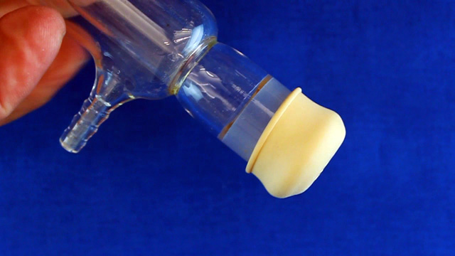
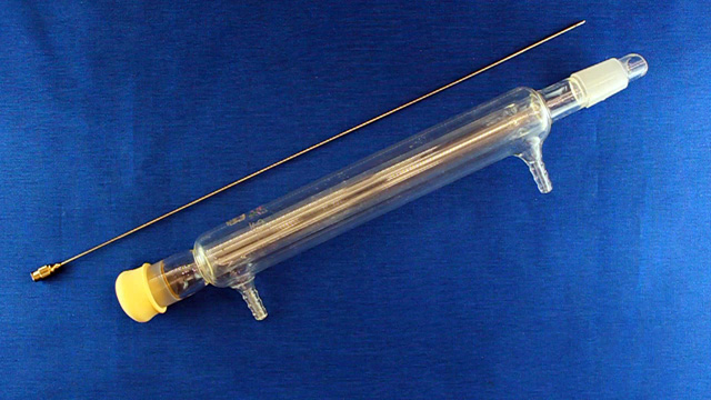
Use a rotating motion to add a teflon sleeve to the end of a condenser.
(Not using a teflon sleeve can result in a permanently polymethylmethacrylate fused joint at the end of the experiment.)
Cap the top of the condenser with a septum.
Insert a long needle through the septum to deliver nitrogen to the bottom of the condenser. Also insert a short needle through the septum for the nitrogen exit.
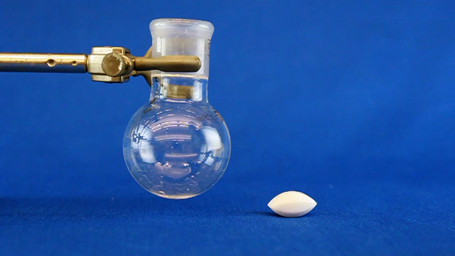
Add a 20x10 mm oval-shaped magnetic stir bar to a 25 mL round bottom flask. (It is important to use a stir bar designed for a round bottom flask for maximum stirring.)
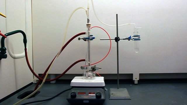
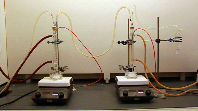
Put a crystallizing dish on a stirrer-hotplate. Clamp the 25 mL round bottom flask in the dish.(Large three-fingered clamps do not work.) Add 16 mL pure water into the round bottom flask and insert a loosely-clamped condenser assembly. Spin the stir bar at the maximum speed that does not cause the stir bar to jump. What is your stir speed?
Connect nitrogen to enter through the long needle and exit through the short needle to a bubbler that monitors flow. Start the nitrogen gas.
(Doing the synthesis without a nitrogen atmosphere produces polydispersed spheres.) Connect the water lines to the condenser (in at the bottom and out at the top).
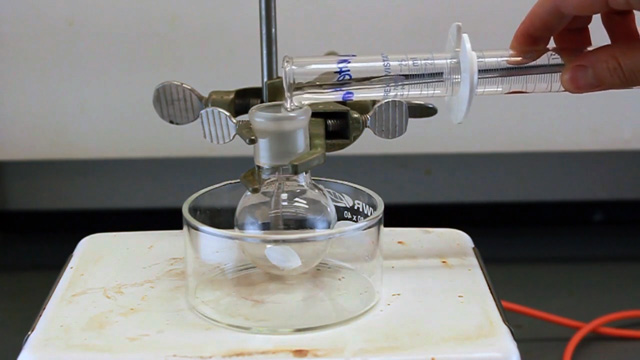
Briefly lift the condenser to add 3.0 mL methyl methacrylate to the round bottom flask. (Using a syringe for addition is best.) The size of the spheres produced depends on temperature, stir rate, and concentration. The methyl methacrylate should form a suspension with uniformly sized spherical drops in the water. If the solution looks clear, check with instructor before continuing. Be sure the round bottom flask is in the center of the hotplate.
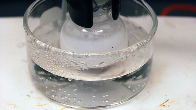
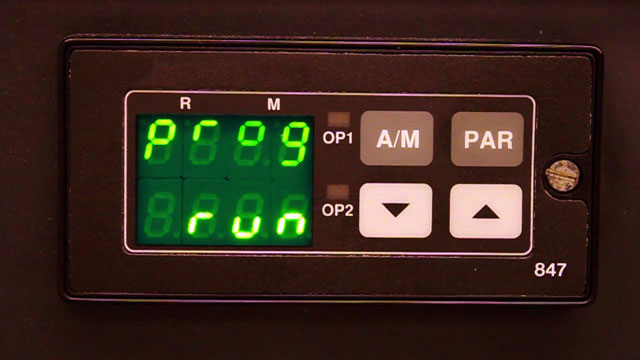
Put water in the crystallizing dish and heat to 70 °C. (An automatic temperature controller may be available.) The crystallizing dish containing water helps minimize temperature fluctuations and provides a place to measure the temperature. Start the water flow through the condensor.
When conditions are stable and an emulsion observable, briefly lift the condenser to quickly add 0.25 to 0.35 mL of 6% 2,2-azobis(2-methyl-propionamidine) dihydrochloride dissolved in water. This compound decomposes with heat to produce a free radical initiator for the polymerization reaction.
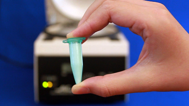
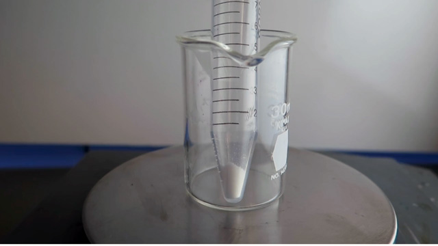
Polymerization is indicated by formation of a milky white suspension after a few minutes. Keep adjusting the temperature to maintain 70 °C for the next 40 minutes.
After 40 minutes of heating, remove the condenser. There should not be a noticeable odor if the polymerization was successful. Be sure to return the Teflon joint sleeve.
Add 500µL of original solution to 20 mL water. If there is time, start the self-assembly experiment. Otherwise store the solution in a large capped centrifuge tube.
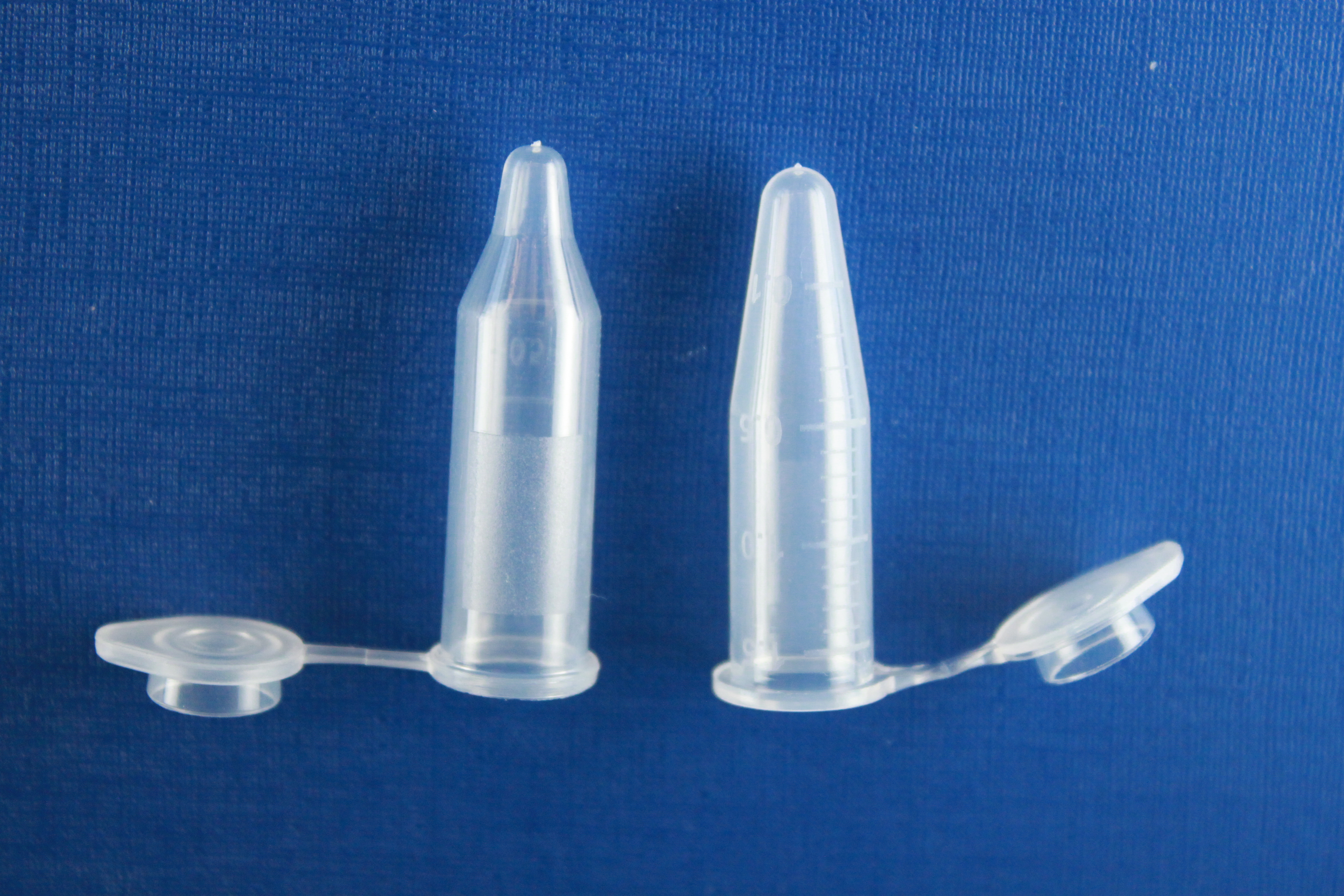
Use the original solution to fill two labeled microcentrifuge tubes to the same depth. Save the capped tubes for the inverse opal synthesis experiments. Ideally these tubes will be centrifuged at 2000 rpm for several hours before the next lab period.
Store any remaining original solution in a capped appropriate-size centrifuge tube for insurance.
Glassware can be cleaned with acetone since acetone dissolves the starting material and the product.
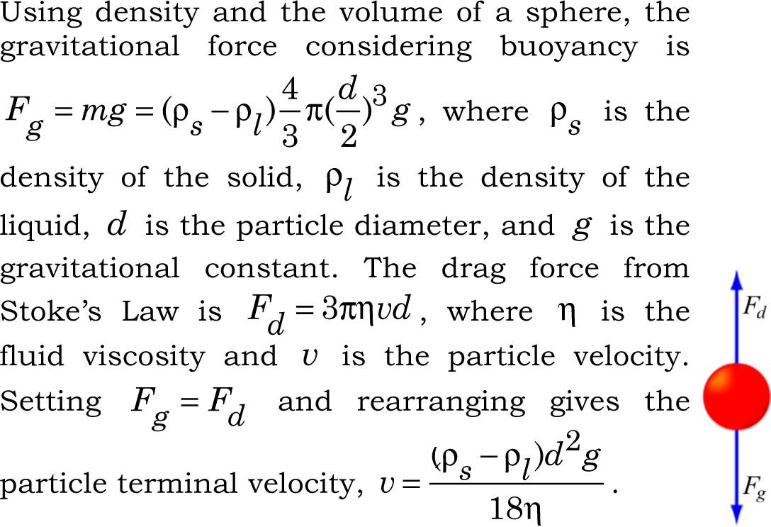
Stoke's Law Determination of Particle Size (Write a purpose and method for the Stoke's Law determination before starting.)
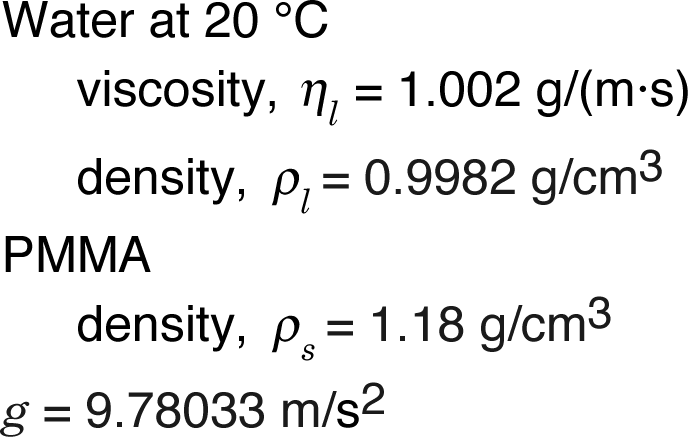
The beads will fall at their terminal velocity, which depends on their size and the known viscoscity and densities. You will measure the velocity to find the particle diameter.
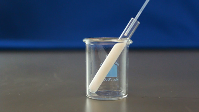
Use a glass Pasteur pipet to fill a small 6x50mm test tube from the bottom up with the polymethylmethacrylate nanosphere suspension. Avoid bubbles.
Seal with stopcock grease so that the water will not evaporate.
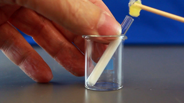
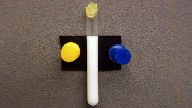
Thumbtack black electrical tape with the sticky side out to a bulletin board. (Use a uniquely colored set of pushpins.)
Align the top of the suspension with the top of the black electrical tape.
Monitor the time and distance between the top of the tape and the settled top.
Record data with a photograph that contains the entire height of the tape so the tape width can be used as a scale bar. Be sure to include the initial data point in your documentation to verify the initial alignment.
Document the sample height every 3-4 days for a month. Upload your photographs to your group folder on the shared drive.
Use ImageJ for measuring lengths in zoomed digital images. (Use the straight line tool to drag the length of the black tape and menu Analyze/Set Scale to tell the program the actual length of the tape, often 19mm. You can then use the straight line tool to measure lengths in real units.) Be sure all measured data is recorded in your laboratory notebook. An alternative is to print each photograph 8.5x11" and use a ruler.
Calculations and Graphs
Update your data table at least weekly. For each data point (total time in seconds, distance fallen in mm), calculate the particle diameter. Is the calculated diameter consistent?
Near the end of the experiment, plot the distance fallen in mm as a function of the number of seconds and use the slope of the linear portion as the best fit velocity. What is the overall velocity in nm/s? What is the particle size based on this velocity? What does it mean if your y-intercept is not zero?
Self-Assembly and Photonic Band Gap Determination of Particle Size (Write a purpose and method for the diffraction experiment before starting. What are the important variables?) This procedure is based on work by William Schreiter, Richard Amankwah and Karen Pearson, Lawrence University (2004).
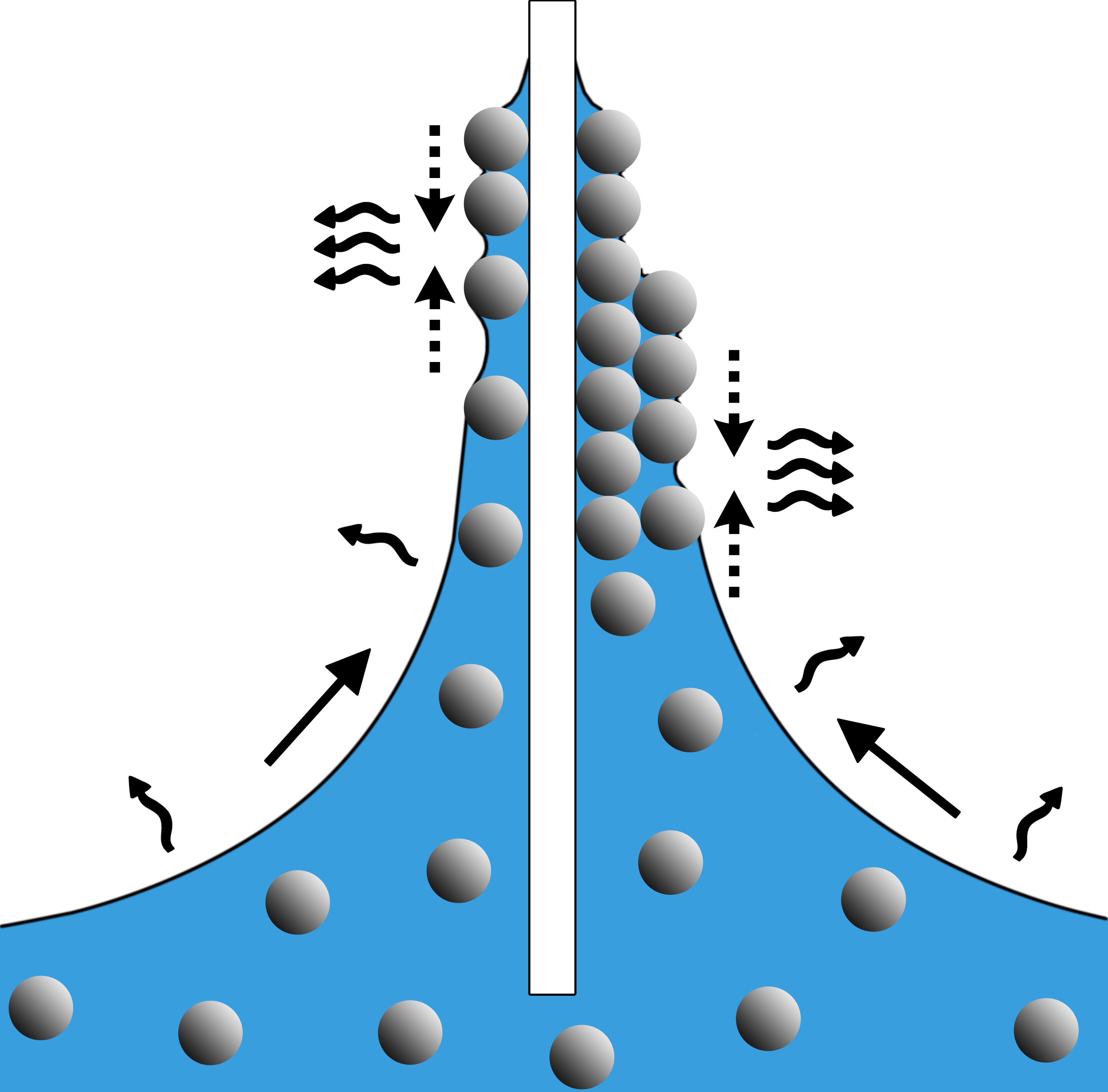
Uniform evaporation of a dilute suspension of polymer nanospheres creates a close packed layer on the surface of a glass slide suspended in the
solution. The particle size can be calculated from the visible absorption maximum (called the photonic band gap) which arises by diffraction from uniformly spaced features.
Dilute 500 microliters of previously prepared PMMA nanosphere solution in
20 mL distilled water. (You probably did this step right after the synthesis.)
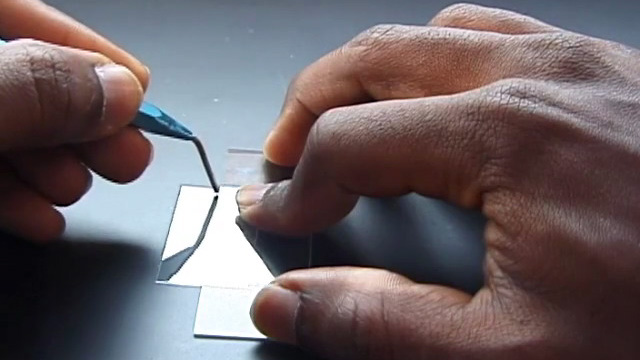
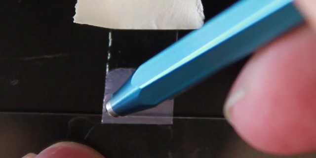
Using a diamond pencil, score and break lengthwise a large glass
cover slip (22 mm x 40 mm). The piece of glass should then be able to fit into the sample compartment for visible spectroscopy measurements.
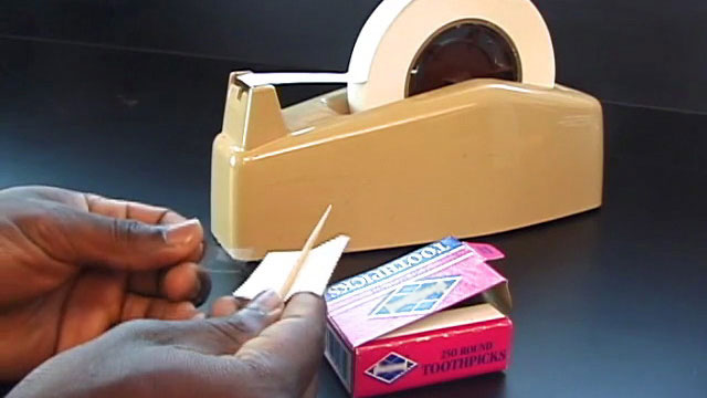
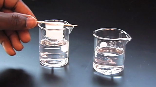
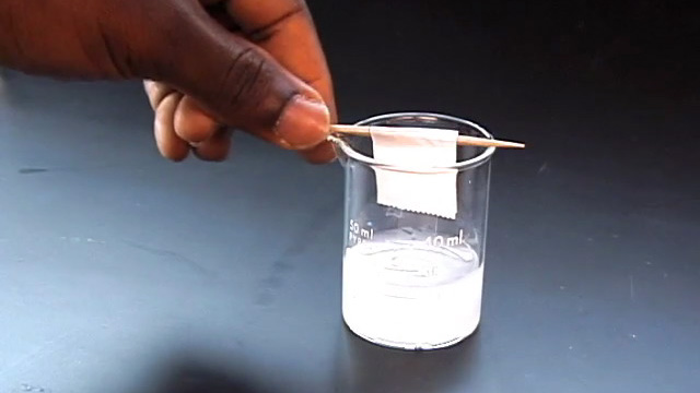
Gently attach one of the glass pieces to a toothpick using tape. Write your name on the tape.
Clean the glass to remove fingerprints and dust by soaking in isopropanol followed by soaking in pure water.
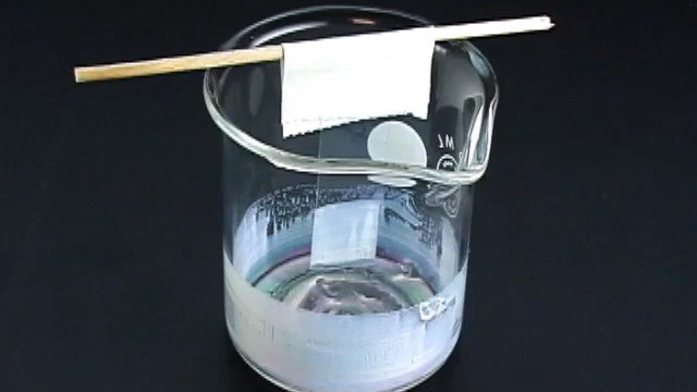
Suspend the glass slide into the diluted PMMA nanosphere suspension you prepared earlier.
If desired, more than one glass slide can be suspended in each solution as long as they do not touch each other.
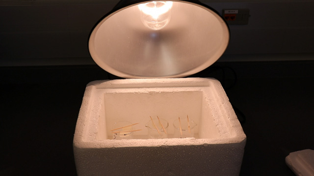
To promote the growth of high quality close packed layers on the glass
slide, evaporate the solution in a controlled temperature environment for several days. Slightly above room temperature works well.
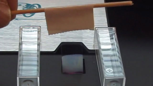
After the solution has evaporated, a dry film of the close-packed nanospheres remains on the glass slide.
Observe the color of the film under reflected light conditions by holding the slide under a bright light source. Observe the color of light transmitted
through the film by holding the glass slide up and looking at a bright light source through the slide. How are the colors of reflected and transmitted light related?
Obtain the visible absorption spectrum when the glass slide is perpendicular to the beam. If an obvious peak is not seen,
repeat the evaporation step and try again with a cleaner slide or more uniform evaporation.
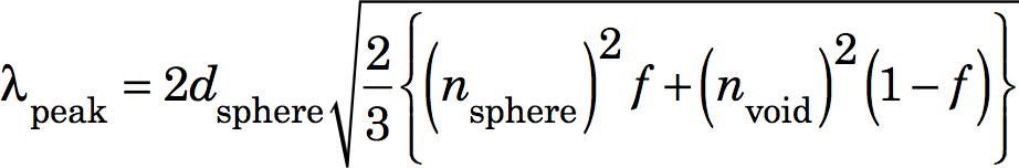
Use a modified Bragg equation to calculate the size of the nanoparticles from the wavelength of the absorbance peak, λpeak. The index of refraction is 1.495 for PMMA and 1.000 for air.
λpeak = wavelength at maximum absorbance (in nm) dsphere = diameter of the close packed spheres (in nm) nsphere = index of refraction for the spheres nvoid = index of refraction for void spaces (presumably air-filled) f = filling fraction for a close packed structure (0.74)
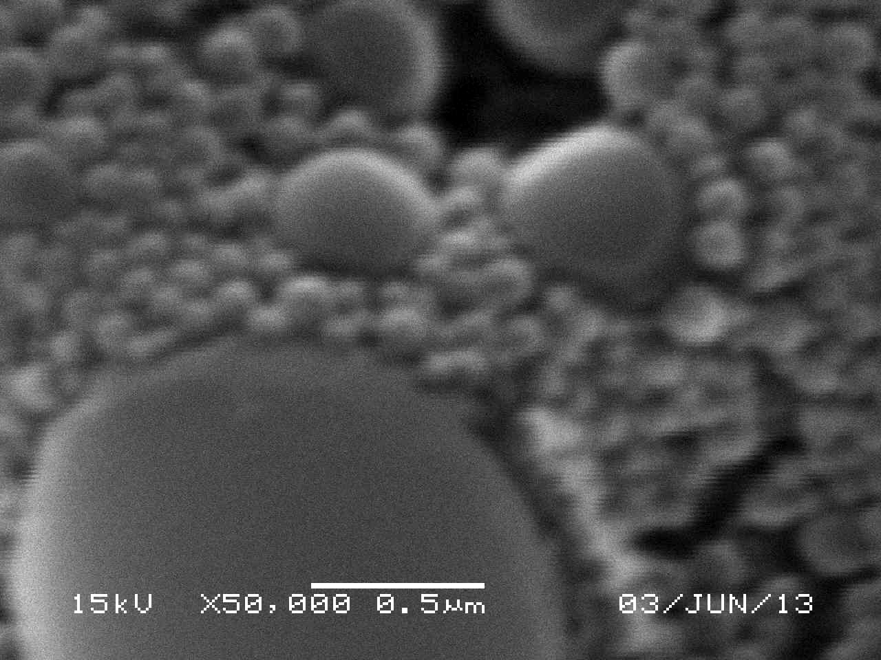
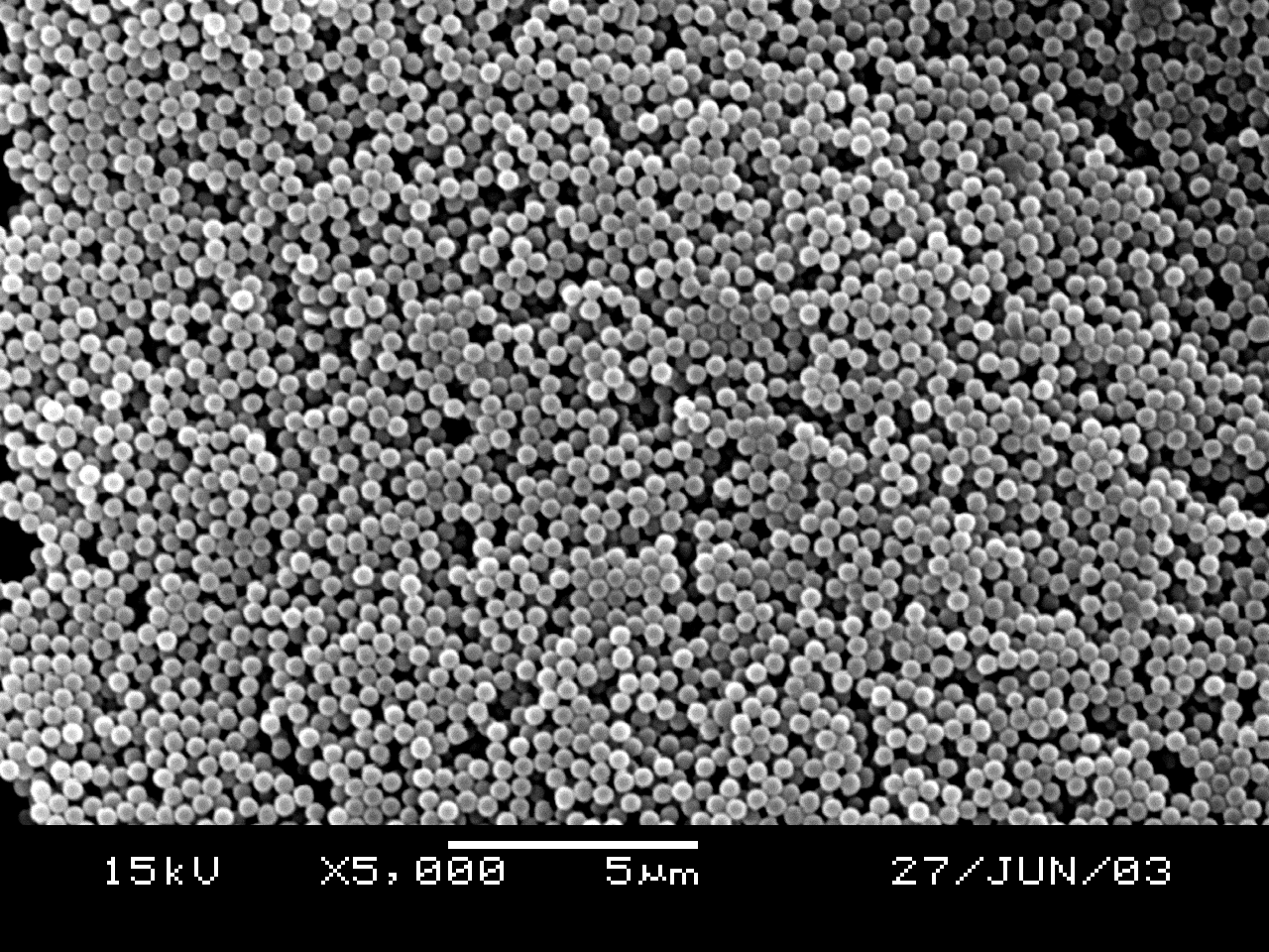
SEM Determination of Particle Size (uses sample from previous step)
Use a diamond pencil to score and then break off a small portion of the cover glass with self-assembled spheres.
Sputter coat and examine by SEM with 10kV and 5000x magnification. Are the spheres close-packed? What size are the spheres? Sphere size is better measured using multiple spheres in a line and then dividing by the number of spheres measured. Does this size agree with your diffraction results? (ImageJ is useful for measuring lengths. Use the straight line tool to drag the length of the scale bar and menu Analyze/Set Scale to tell the program the actual length of the scale bar. You can then use the straight line tool to measure lengths in real units.)
Synthesis of Inverse Opal Photonic Crystals (Write a purpose and method for the synthesis before starting. What is the chemical reaction?) This procedure is derived from the Inverse Opal Photonic Crystals Laboratory Guide by R. Schroden and
N. Balakrishnan, University of Minnesota MRSEC, 2001.
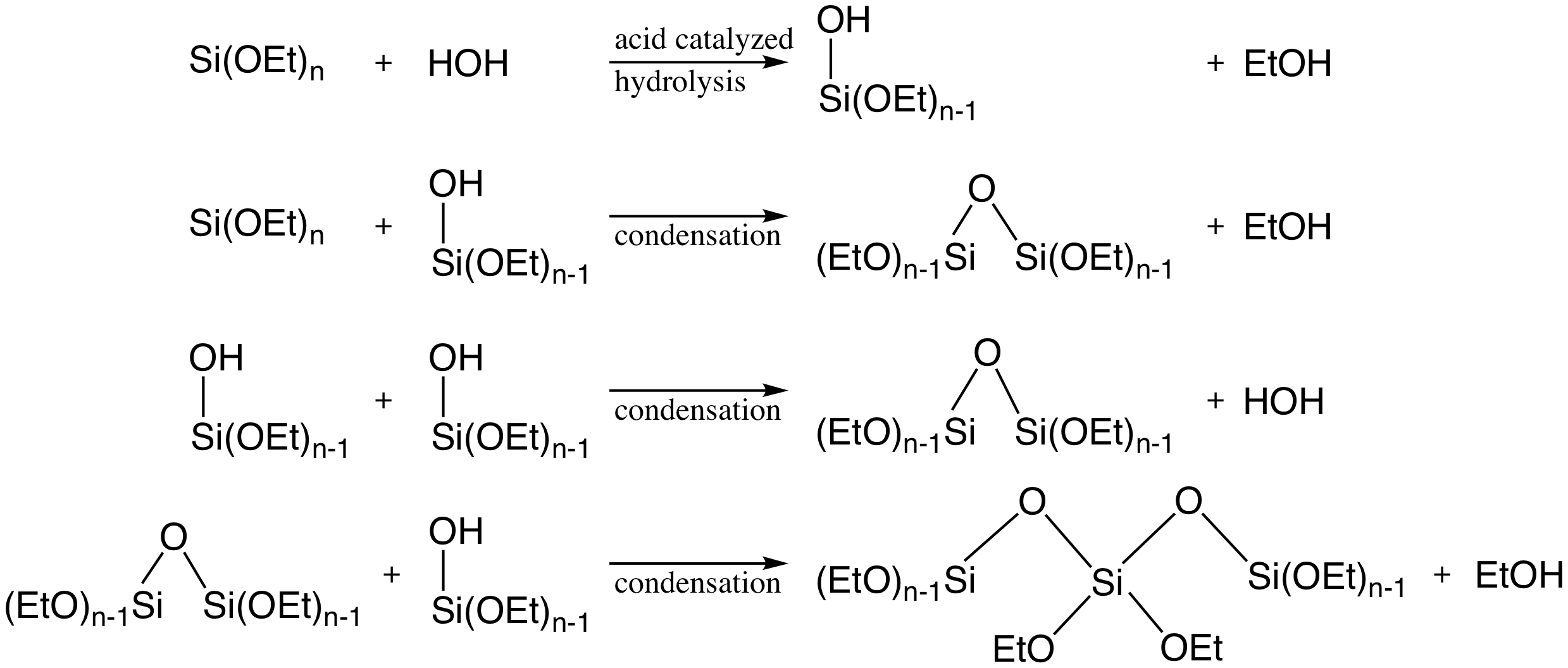
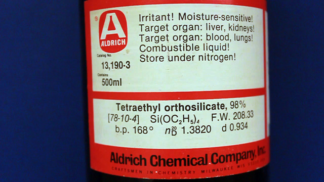
A Stöber synthesis using tetraethylorthosilicate produces a silica solid by a condensation polymerization.
Forming the silica in the spaces between close-packed polymethylmethacrylate spheres used as a template, followed by removal of the polymethylmethacrylate spheres through heating, yields a dimensionally ordered arrangment of holes in the silica solid.
This photonic material will change color if the index of refraction of the material within the pores is changed.
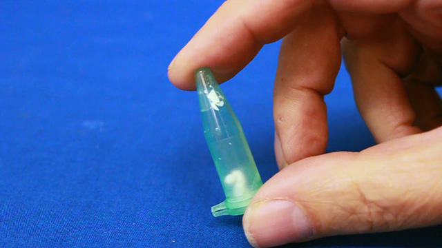
Transfer a portion of your sample to two 1.7 mL microcentrifuge tubes. Always balance the centrifuge
by pairing tubes such that opposing tubes have the same amount of solution. Spin your sample at 2000 rpm for several hours.
(Spinning at 5000 rpm does not pack as well and spinning at 10,000 rpm will crush the product.)
It is desirable to spin the samples before the lab period starts or the experiment will take an extra period.
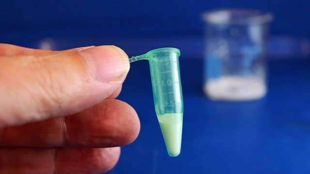
Remove and discard the water above the polymerized polymethylmethacrylate spheres. Caution: Squeeze the dropper before immersing in the water, lower the dropper to just above the solid, and then release to remove the water. If you squeeze with the dropper in the water you will need to repeat the centrifuge step. Put the liquid in the organic waste.
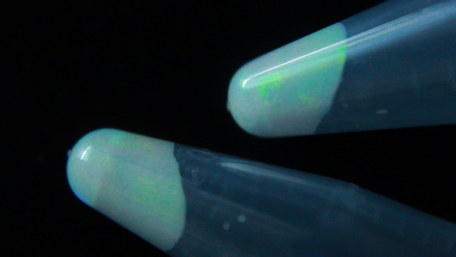
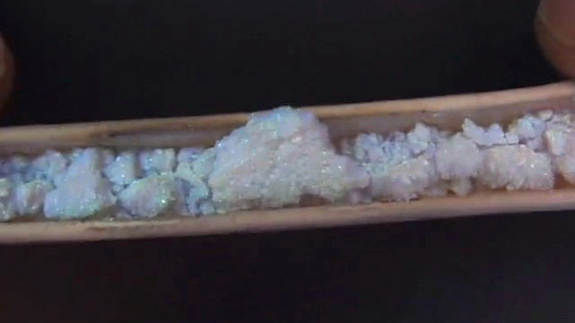
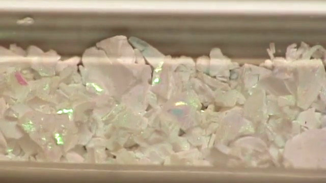
Successful product will change colors with the angle of observation or lighting; the product should look iridescent.
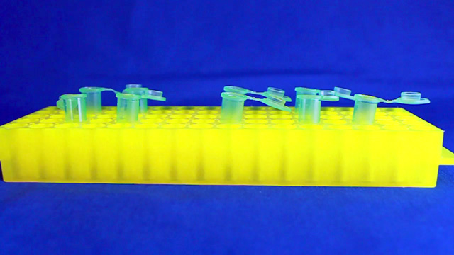
Leave the lid open and store the tube in a rack to dry for week.
Dry the sample until it can be dumped from the tube.
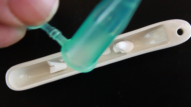
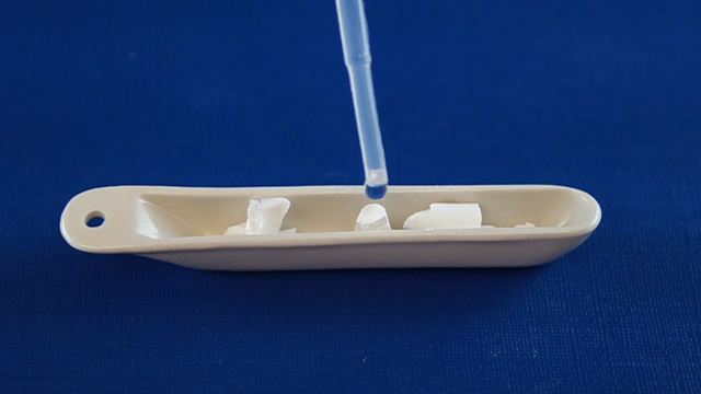
Pour the solid into a combustion boat.
Prepare a well-mixed solution of 1.0 mL 100% ethanol, 1.5 mL tetraethylorthosilicate, 1.0 mL 25% hydrochloric acid. (This is enough for many samples but it must be freshly prepared.)
Wet the spheres with the solution but once all the spheres are wet stop adding liquid or the product may be too dense.
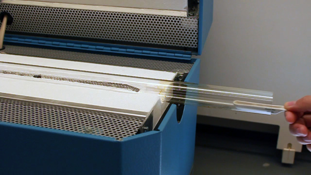
Place the combustion boats into the quartz liner of a tube furnace (or a ventilated box furnace) in a hood.
Ramp the temperature at 2 °C/minute from room temperature to 300 °C to complete the silica formation. Hold at 300 °C for 2 hours. Ramp the temperature at
2 °C/minute to 550 °C to decompose the polymethylmethacrylate spheres. Hold at 550 °C overnight (ten hours). Cool the oven to room temperature.
High quality samples will be apparent by their opalescence.
SEM Determination of Inverse Opal Hole Size (uses sample from previous step)
Stick a portion of your sample to the carbon tape on the SEM holder and add your name to the sample map. Samples will then need to be sputtered since SiO2
is non-conducting. On our instrument (using 25kV with spotsize 27 and z 15mm) it should be possible to see holes at 3000x. Look carefully.
Hole size is better measured using multiple holes in a line and then dividing by the number of holes measured.
(ImageJ is useful for measuring lengths. Use the straight line tool to drag the length of the scale bar and menu Analyze/Set Scale
to tell the program the actual length of the scale bar. You can then use the straight line tool to measure lengths in real units.) Be sure to include details of your
analysis in your laboratory notebook.
Inverse Opal Properties (uses sample from previous step)
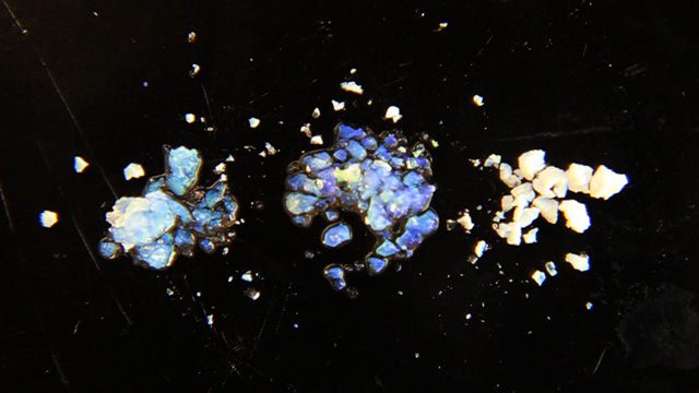
What color is your product in air? (The index of refraction of silica is 1.460 and of air is 1.000)
Add a drop of ethanol to a small portion of your product. What color do you observe? (The index of refraction of ethanol is 1.360)
Add a drop of toluene to a small portion of your product. What color do you observe? (The index of refraction of toluene is 1.496)
Spectra, Images and Graphs
Your notebook should include your Stoke's Law photographs and graph, optical diffraction spectra, and SEM images of both opal and inverse opal materials.
Conclusions
Did your methyl methacrylate polymerize? What observations support your answer? Using your chemical knowledge of polar/nonpolar, physical properties, and the polymerization mechanism, explain why solid spheres are expected to be produced.
Based on your Stoke's Law measurements, what is the velocity (nanometers/second) and the corresponding diameter (nanometers) of the spheres?
Based on evaporative self-assembly to pack spheres, what is value for the photonic band gap (nanometers or eV) and the corresponding diameter (nanometers) of the spheres? What did you use to obtain a constant temperature? How many times did you try the evaporation?
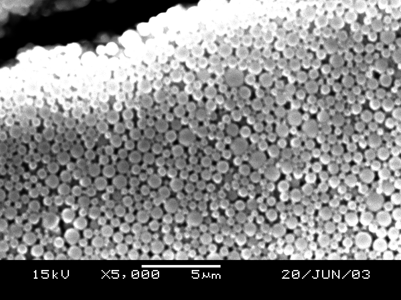
Based on the SEM image of the opal, did the spheres close-pack and what is the diameter of the spheres (nanometers)? You should measure the diameter for more than one sphere and more than one image and take an average. Report all measured data.
Based on the SEM image of the inverse opal, are the holes close-packed and what is the diameter of the holes (nanometers)? You should measure the diameter for more than one hole and more than one image and take an average. Report all measured data.
Based on your hole diameter, predict the absorption wavelength for air, ethanol, and toluene. (How do you need to modify the equation for closed-packed spheres for the inverse material?) How well do your predictions match the observed colors?
What size are the nanospheres? Do the different methods give the same results within experimental error?
Materials for Synthesis and Stokes Law
More than one apparatus can use the same nitrogen supply, as long as they are run in parallel or started in order with the upstream apparatus sealed before the next is started. We routinely use four setups in series per hood.
Stirrer/hotplate. A Corning PC-420D with 6795PR temperature controller is convenient.
25 mL round bottom flask, condenser, crystallizing dish
Developed in collaboration with the University of Wisconsin Materials Research Science and Engineering Center Interdisciplinary Education Group | UW MRSEC This page created by George Lisensky, Beloit College.
Last modified February 6, 2020.
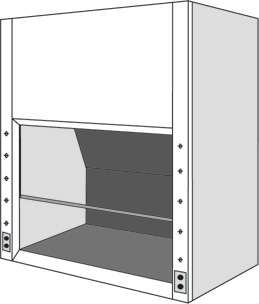 Fumehood required
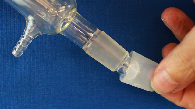
 Click image for larger view
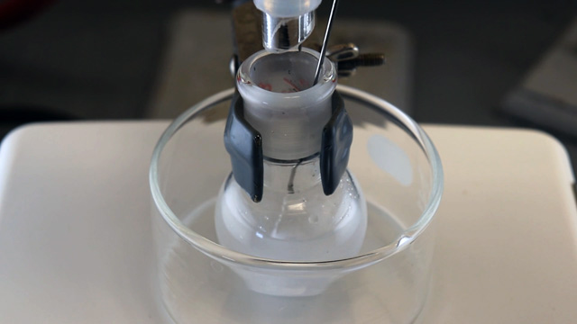
 Click image for larger view
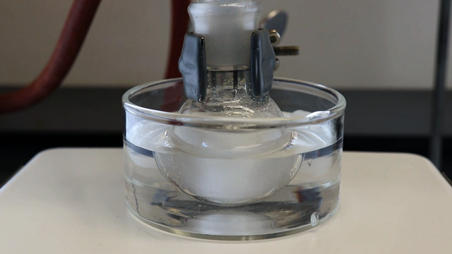
 Click image for larger view
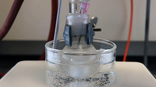
 Click image for larger view
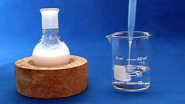

 Click image for larger view