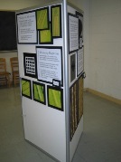
 Probe Microscopy Exhibit
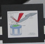
Probe microscopy is a valuable tool in nanotechnology. Objects on the nanoscale are too small to be seen by the naked eye and most optical microscopes. Probe microscopy utilizes the interactions between a sharp probe tip and a nanoscale object in order to measure the object's properties, including size, shape, stiffness, and electrical and magnetic characteristics.
Measuring Magnetism
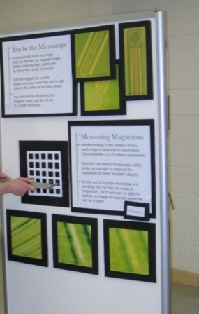
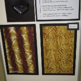
The magnetic force is one type of force that probe microscopes use in order to image an object. On this panel of the exhibit, visitors learn about how magnetism produced the images of the DVD Rom disk seen along the bottom. (Images courtesy of Dr. Charles Paulson and Prof. Dan van der Weide)
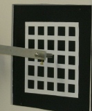
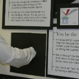
The visitor is invited to "become the microscope" and find the pattern behind the black panel by feeling the magnetic interaction of the probe and the material.

Scoping Out Stiffness
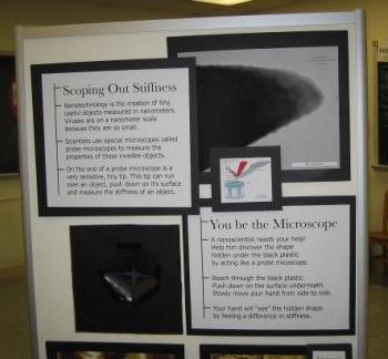 Images can also be created by pushing on a nanoscale surface with a
probe tip and mapping out areas that are hard or soft. The visitor can
again "become the microscope" by placing his or her hand inside the black
box and discerning the patterns based on where he or she feels hard or soft
material beneath his or her fingertips. This type of microscopy can be used to
measure soft biological nanomaterials, like the tobacco mosaic virus, seen
below. (Image courtesy of Matt D'Amato and Prof. Rob
Carpick)
Mapping Mini Mountains
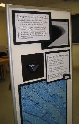 This panel of the exhibit teaches the visitor how the surface of a
nanomaterial can be imaged using probe microscopy. This microscopy is so
sensitive it can even measure a single atom on a surface! The image at the
bottom of the panel shows the visitor individual silicon atoms on a surface. (Image courtesy of Prof. Max Lagally) By placing
a hand inside the black box, the visitor can "become the microscope" and feel
a pattern delineated by bumps in the unseen surface.

