
Atomic Force Microscopy
Introduction
Could you pick up a boulder with a pair of tweezers? Could you pick up a piece of hair with a bulldozer? Having the right size tools for the job can be pretty important. When scientists study the nanoscale, they need to use tools that are the right size. To study things that are very, very small, they need very, very small tools.
Scanning probe microscopes are a set of tools with very small parts that help scientists image the nanoscale. Each type of scanning probe microscope involves a very fine probe tip that scans back and forth over a surface. The first SPM was the scanning tunneling microscope (STM). STM is limited to surfaces that are electrically conductive. Later, Atomic Force Microscopy (AFM) was invented, which could scan any surface, conductive or not.
How does AFM Work?
AFM works by bringing an atomically sharp tip close to a surface. There is an attractive force between the tip and the surface and this force is kept the same throughout the experiment. As the probe tip scans back and forth over the surface, the tip will rise and fall with the different features on the surface. Since all this is going on at a very small scale, we can't watch the tip directly. A laser is pointed at the tip and is reflected to a sensor. As the tip goes up and down the laser hits different parts of the sensor. With the information the sensor collects, an image of the surface can be recreated.
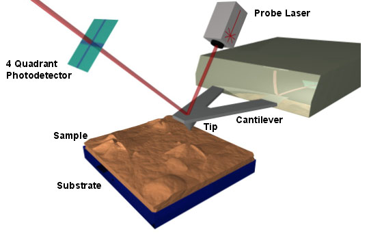
AFM - a super sharp tip moves across a sample; as the tip moves up and down over the surface, a laser detector records the height.1
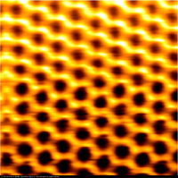
An AFM image of graphite. The image size is 2 x 2 nanometers.2
References
1. Image from www.barrett-group.mcgill.ca
2. Image from www.physik.uni-augsburg.de