
![]()
Introduction
In 1952, Bloch and Purcell won the Nobel Prize in Physics for the development of the analytical technique known as nuclear magnetic resonance (NMR). Magnetic resonance imaging (MRI) is an adaptation of NMR for medical diagnostic purposes.
Both NMR and MRI rely on the fact that some atomic nuclei have a magnetic moment associated with their spinning. The most common nuclei studied using NMR are H-1, C-13, F-19 and P-31. Most MRI studies involve the H-1 nuclei in water.
The isotopes listed above have two spin states. When these nuclei are exposed to an external magnetic field, their spins can be parallel or anti-parallel to this external field. Transitions between these two spin states can be induced with energy from radio waves of the proper frequency. A radio frequency (rf) signal is transmitted to the sample, which has been placed in a strong magnetic field.
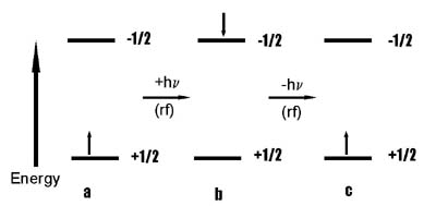
A slight excess of the nuclei of the atoms are initally in the lower energy state, represented by a above. The nuclei in the sample interact with the rf signal. This interaction causes the spins of the nuclei, represented by a, to flip to the less stable direction of the higher energy state represented by b. The nuclei relax to the more stable state through the emission of a rf signal of the same frequency. This is represented above by going from b to c. A rf receiver measures the signal after its interaction with the sample. The analysis of these measurements can help to identify the structure of the sample, because the relaxation times for the nuclei are dependent upon their chemical environment.